The Human Visual System#
Further Reading
This page provides a high-level overview of the human visual system, but it is not intended as a thorough treatment of the subject. For further reading, we suggest The Foundations of Vision by Brian Wandell [Wandell, 1995], available online here.
The human visual system begins in the eye where light passes through the optics of the front of the eye and through to the retina. In the retina, various photoreceptors detect the light and pass signals through mediating cells to the first neurons of the visual pathway called retinal ganglion cells (RGCs), which connect directly to the brain. The visual field is transformed but topologically preserved on the retina (Fig. 1), and functional regions of the brain in which this topological organization is preserved are called “retinotopic maps”.
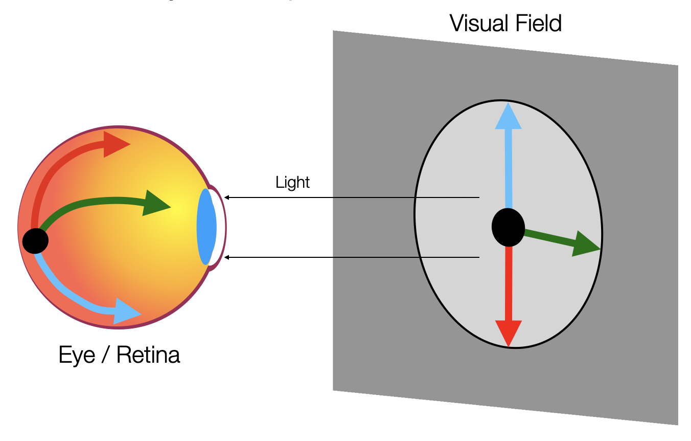
Figure 1. Light from the visual field passes through the optics of the eye to the retina. On the back of the retina, the neurons that process the visual field form a transformed but topologically preserved map of it. Such topologically preserved maps in the brain are called “retinotopic maps”.#
Although vision begins in the eyes, and although substantial visual processing begins in that very first layer of mediating cells, most of human vision happens in the brain.
The first post-retinal synapse (i.e., junction between neurons) of the visual system is in a subcortical brain structure called the lateral geniculate nucleus (LGN). RGCs from the retina are routed through a junction near the front of the brain called the optic chiasm where they are sorted according to whether they receive input from the left or the right half of the visual field. Neurons from both eyes that receive input from the right half of the visual field project through the optic chiasm to the brain’s left hemisphere, and neurons receiving input from the left visual field project to the right hemisphere (Fig. 2).
“subcortical”
A “subcortical” brain structure is a structure that is buried beneath the cerebral cortex. The cerebral cortex, often just called cortex, is a thin layer covering the large wrinkled part of the top of the brain and is made of the brain’s “gray matter”, which surrounds the subcortical “white matter”. Brain structures like the amygdala and the cerebellum are also subcortical. Cortical brain regions include Broca’s area and the motor homunculus. The white matter and gray matter of a hemisphere together are sometimes called a cerebral hemisphere.
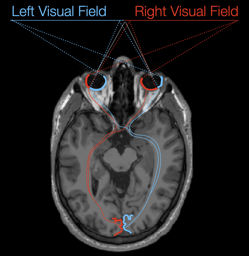
Figure 2. The left and right visual field project through the optics of the eye onto particular parts of the retina, no matter where the eyes are pointed. The RGC neurons of the retina project through a junction called the optic chiasm where they are sorted based on the half (left or right) of the visual field from which they receive input, and back to the LGN (not shown) of the contralateral hemisphere (right visual field projects to left hemisphere and left to right). In the LGN, the RGCs synapse with neurons whose cell bodies lie in the primary visual cortex (V1), at the very back of the brain.#
These neurons that connect the retina to the visual cortex in the back of the brain run along fiber tracts in the brain’s “white matter”. In Figure 2, there is a thin layer of darker tissue visible along the edge of the hemispheres surrounding slightly lighter tissue. The lighter tissue in the middle of the brain is the white matter, and it consists primarily of connections between neurons, a bit like the wires that connect the processors of a supercomputer. The darker tissue surrounding the white matter is called the “gray matter” and makes up the cerebral cortex. It contains most of the cell bodies of the neurons in the cerebrum, much like the processors of a supercomputer themselves.
The gray mater consists of several thin layers that are topologically equivalent to the (2D) surfaces of spheres. This 2D shape is important because it means that we can visualize and describe regions of the brain in terms of where they lie on a 2D surface instead of in terms of a 3D space, and, in fact, we often inflate the surface of hemisphere of a surface to aid in visualization (Fig. 3). For the purposes of this project, we will ignore all functional differences between layers of the gray matter and treat it as purely 2D.
Are the hemispheres really topologically spheres?
Arguably, the cortical surface of a hemisphere of the brain isn’t really topologically equivalent to a sphere because the corpus callosum makes a hole in the sphere along the hemisphere’s medial surface. This is perfectly true, but we typically treat the medial surface as a very thin surface of gray matter in order to make calculations like brain inflation simpler.
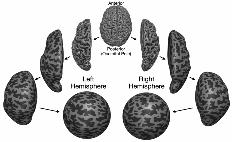
Figure 3. Each hemisphere of the brain can be separately inflated into a sphere by iteratively smoothing the cortical surface. Once the surface has been inflated to a sphere, an orthographic projection of the sphere can be used to make a 2D disk called a “flatmap” that represents part of the cortical surface. These flatmaps are often useful for visualization. In the figure, the dark gray indicates sulci (valleys) on the cortical surface while the light gray indicates gyri (peaks).#
The 2D surface of the human visual cortex is tiled with separate functional regions (i.e., patches of cortex that perform a similar function). The first of these regions is called “V1” as well as “primary visual cortex” and “striate cortex”. V1 is the location where neurons from the LGN first arrive at cortex, and it is responsible in part for processing lines and edges in the visual field [Hubel and Wiesel, 1959]. From V1, neurons project to many other visual areas, most of which cover the posterior part of the cerebral cortex (Fig. 4). Together, these regions of the visual cortex makes up approximately 25% of the cortical surface in humans.
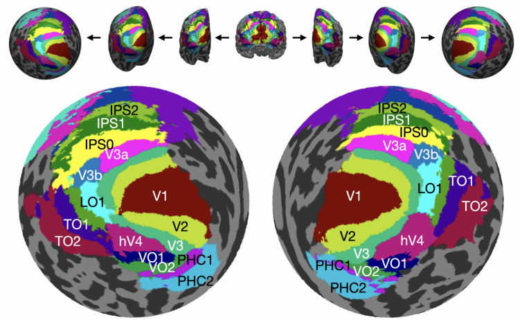
Figure 4. Flattened orthographic projections of the posterior left and right cortical surfaces of an example subject. In each map, numerous visual areas are colored and labeled based on the Wang full probability atlas of visual areas [Wang et al., 2015].#